Home
/ Tendons And Ligaments In Foot And Leg - 8 Foot Pictures Ideas Ankle Anatomy Foot Anatomy Foot Pictures - The tendon passes behind the inner ankle bone (medial malleolus) and underneath the foot attaching to the tarsal bones.
Tendons And Ligaments In Foot And Leg - 8 Foot Pictures Ideas Ankle Anatomy Foot Anatomy Foot Pictures - The tendon passes behind the inner ankle bone (medial malleolus) and underneath the foot attaching to the tarsal bones.
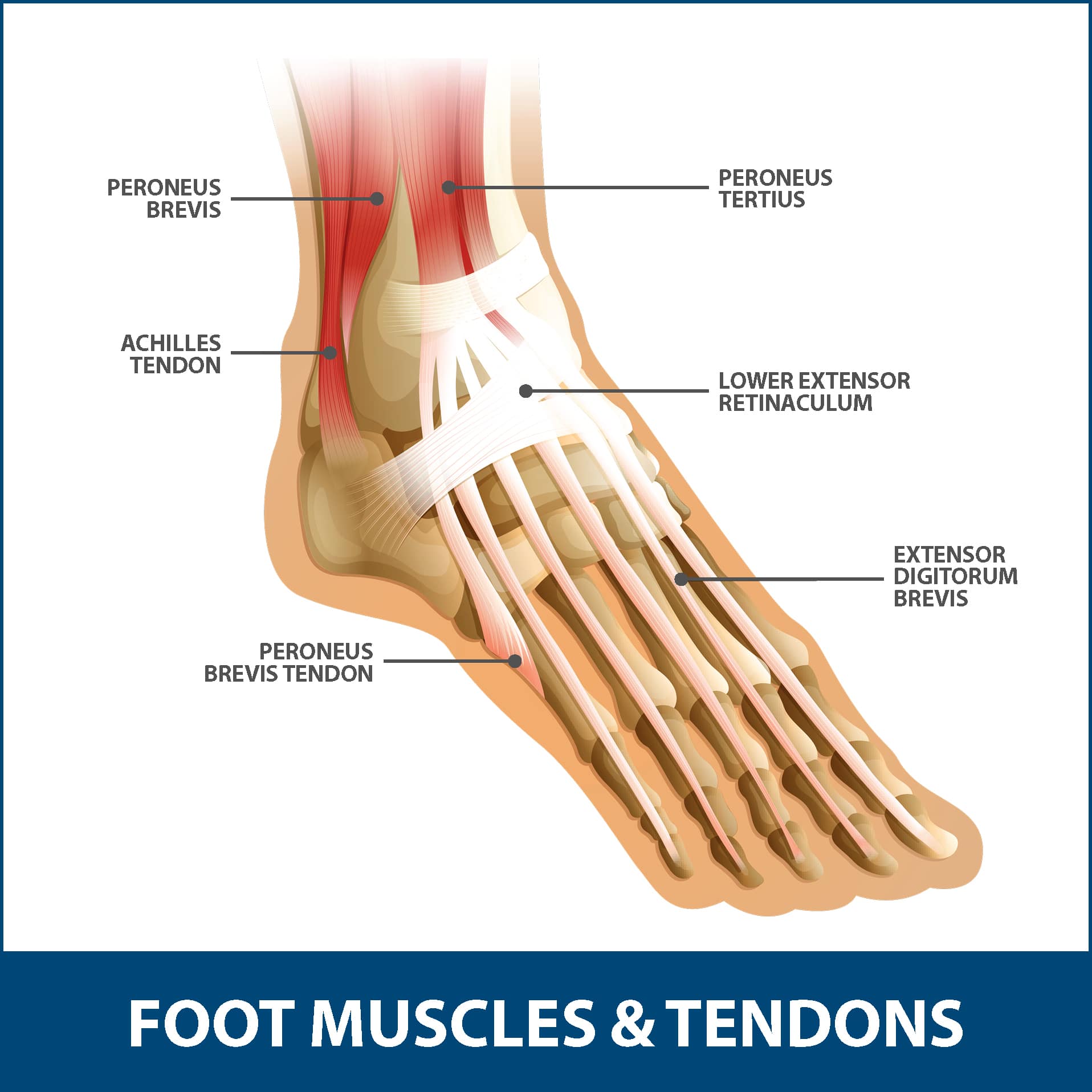
Tendons And Ligaments In Foot And Leg - 8 Foot Pictures Ideas Ankle Anatomy Foot Anatomy Foot Pictures - The tendon passes behind the inner ankle bone (medial malleolus) and underneath the foot attaching to the tarsal bones.. The peroneus brevis tendon inserts into a tuberosity at the base of the fifth metatarsal bone, on its lateral side. Ligaments and tendons are both made up of fibrous connective tissue, but that's about where the similarity ends. The foot is not only complicated in terms of the number and structure of bones, but also in terms of its joints. Tendons can tear partially or completely during a joint injury. It consists of 28 bones, which can be divided functionally into three groups, referred to as the tarsus, metatarsus and phalanges.
While tendons connect muscle to bone, ligaments connect bones to other bones. The ligament, located in the center of the knee, that controls rotation. The four main ligaments in the knee connect the femur (thighbone) to the tibia (shin bone), and include the following: (heel bone) is the largest bone in the foot. Ligament tears are most common for the lateral ligament complex, which include the anterior talofibular (atfl), the calcaneofibular (cfl), and posterior talofibular (ptfl) ligaments.
Ankle Ligament Injuries Podiatry Orthopedics Physical Therapy from www.southfloridasportsmedicine.com #muscle and tendon pain in legs #muscles and tendons of the leg and foot #muscles and tendons of the lower leg #muscles ligaments and tendons of the lower leg #muscles tendons and ligaments of the upper leg It's also instrumental in bending the knee. This is the longest ligament in the foot. It consists of 28 bones, which can be divided functionally into three groups, referred to as the tarsus, metatarsus and phalanges. The peroneus muscles plantarflex and everts the foot. Ligament tears are most common for the lateral ligament complex, which include the anterior talofibular (atfl), the calcaneofibular (cfl), and posterior talofibular (ptfl) ligaments. The ligaments of the foot and ankle can be divided into groups including: In contrast, a ligament consists of bands of thick connective tissue that join bone to bone.
Tendons are long thin bands that attach your muscles to bones.
For example, knee ligaments connect your thighbone to your shinbone, forming a joint, which lets. 2 your feet contain more than 100 muscles, tendons, and ligaments. It consists of 28 bones, which can be divided functionally into three groups, referred to as the tarsus, metatarsus and phalanges. The tibialis posterior tendon is the main invertor of the foot and also helps the calf muscles to plantarflex the foot. Ligaments and tendons are both made up of fibrous connective tissue, but that's about where the similarity ends. Ligaments and tendons are both made up of fibrous connective tissue, but that's about where the similarity ends. Ligament tears are most common for the lateral ligament complex, which include the anterior talofibular (atfl), the calcaneofibular (cfl), and posterior talofibular (ptfl) ligaments. Healthy tendons and ligaments for movement to occur, skeletal muscle must contract but they need the help of tendons and ligaments. Tendons and ligaments in foot and leg / extensor tendons of foot illustration stock image c047 6014 science photo library : Drop that leg back down and swing the other leg up, taking another step forward. Tendons are tough, connective tissue that connects a skeletal muscle to a bone. A ligament is fibrous tissue that connects 2 or more bones together. Tendonitis is an inflammation surrounding a tendon.
When a ligament tears, the resulting injury is often referred to as a sprain. Ligaments connect bones to each other to support a joint. The calf muscle typically gets strained when the foot suddenly bends upward, stretching the calf muscle beyond its limits. The foot is a complex structure that consists of 26 bones, 33 joints, and over 100 muscles, tendons, and ligaments. It involves the distal tibiofibular syndesmotic ligaments.
Foot Anatomy Bones Ligaments Muscles Tendons Arches And Skin from biologydictionary.net Ligaments are the strong and flexible tissues that hold the bones throughout your body together; The main ligaments of the foot include the: The anterior talofibular ligament (atfl), which connects the front of the talus bone to a long bone in the lower leg called the fibula; The foot is not only complicated in terms of the number and structure of bones, but also in terms of its joints. Tendons can tear partially or completely during a joint injury. A high ankle sprain is less common in everyday life but can be seen in competitive athletes. The calcaneofibular ligament (cfl), which connects the calcaneus, or heel bone, to the fibula The tendon passes behind the inner ankle bone (medial malleolus) and underneath the foot attaching to the tarsal bones.
A tendon connects muscle to bone.
The peroneus muscles plantarflex and everts the foot. Ligaments and tendons are both made up of fibrous connective tissue, but that's about where the similarity ends. It allows your foot to flex as you walk or run. The ligament, located in the center of the knee, that controls rotation. The four main ligaments in the knee connect the femur (thighbone) to the tibia (shin bone), and include the following: Reach your arms straight forward and try to hit your arm with your toes. Two muscles make up the calves of the lower leg. Ligaments are elastic bands of tissue that connect bones to each other and provide stability and strength to the joint. The medial deltoid ligament is injured less often. The soleus muscle lies underneath the gastrocnemius. The tibialis posterior tendon is the main invertor of the foot and also helps the calf muscles to plantarflex the foot. The interosseous membrane the interosseous membrane is composed of strong fibrous tissue and runs along the tibia and fibula, and keeps the two bones moving as one unit (figure 4). Ligaments are strong bands of tissue that connect bones to each other and keep your joints stable.
Calf muscle strains are common in athletes, especially tennis players and joggers. Healthy tendons and ligaments for movement to occur, skeletal muscle must contract but they need the help of tendons and ligaments. Drop that leg back down and swing the other leg up, taking another step forward. Ligaments are very similar to tendons. It consists of 28 bones, which can be divided functionally into three groups, referred to as the tarsus, metatarsus and phalanges.
Achilles Tendon Rupture Info Florida Orthopaedic Institute from www.floridaortho.com The calcaneofibular ligament (cfl), which connects the calcaneus, or heel bone, to the fibula Because they are so complicated, human feet can be especially prone to injury. The anterior talofibular ligament (atfl), which connects the front of the talus bone to a long bone in the lower leg called the fibula; A tendon connects muscle to bone. A high ankle sprain is less common in everyday life but can be seen in competitive athletes. For example, knee ligaments connect your thighbone to your shinbone, forming a joint, which lets. The calf muscle typically gets strained when the foot suddenly bends upward, stretching the calf muscle beyond its limits. The ligaments of the foot and ankle can be divided into groups including:
Its unique design allows the foot to handle hundreds of tons of force every day.
Depending on the severity of the injury, treatment by an orthopedic surgeon may be required to treat torn ligaments in the foot. The four main ligaments in the knee connect the femur (thighbone) to the tibia (shin bone), and include the following: The peroneus longus tendon then continues in a plantar direction along the sole of the foot to the base of the first metatarsal bone. For example, knee ligaments connect your thighbone to your shinbone, forming a joint, which lets. Tibialis posterior is the deepest muscle on the back of the leg. Drop that leg back down and swing the other leg up, taking another step forward. The anterior talofibular ligament (atfl), which connects the front of the talus bone to a long bone in the lower leg called the fibula; The talus bone supports the leg bones (tibia and fibula), forming the ankle. The ligament, located in the center of the knee, that controls rotation. The ligaments of the foot and ankle can be divided into groups including: Tendons are tough, connective tissue that connects a skeletal muscle to a bone. The tibialis posterior tendon is the main invertor of the foot and also helps the calf muscles to plantarflex the foot. A tendon connects muscle to bone.

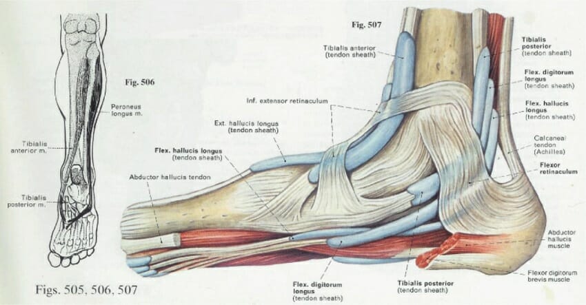
 and underneath the foot attaching to the tarsal bones.)